
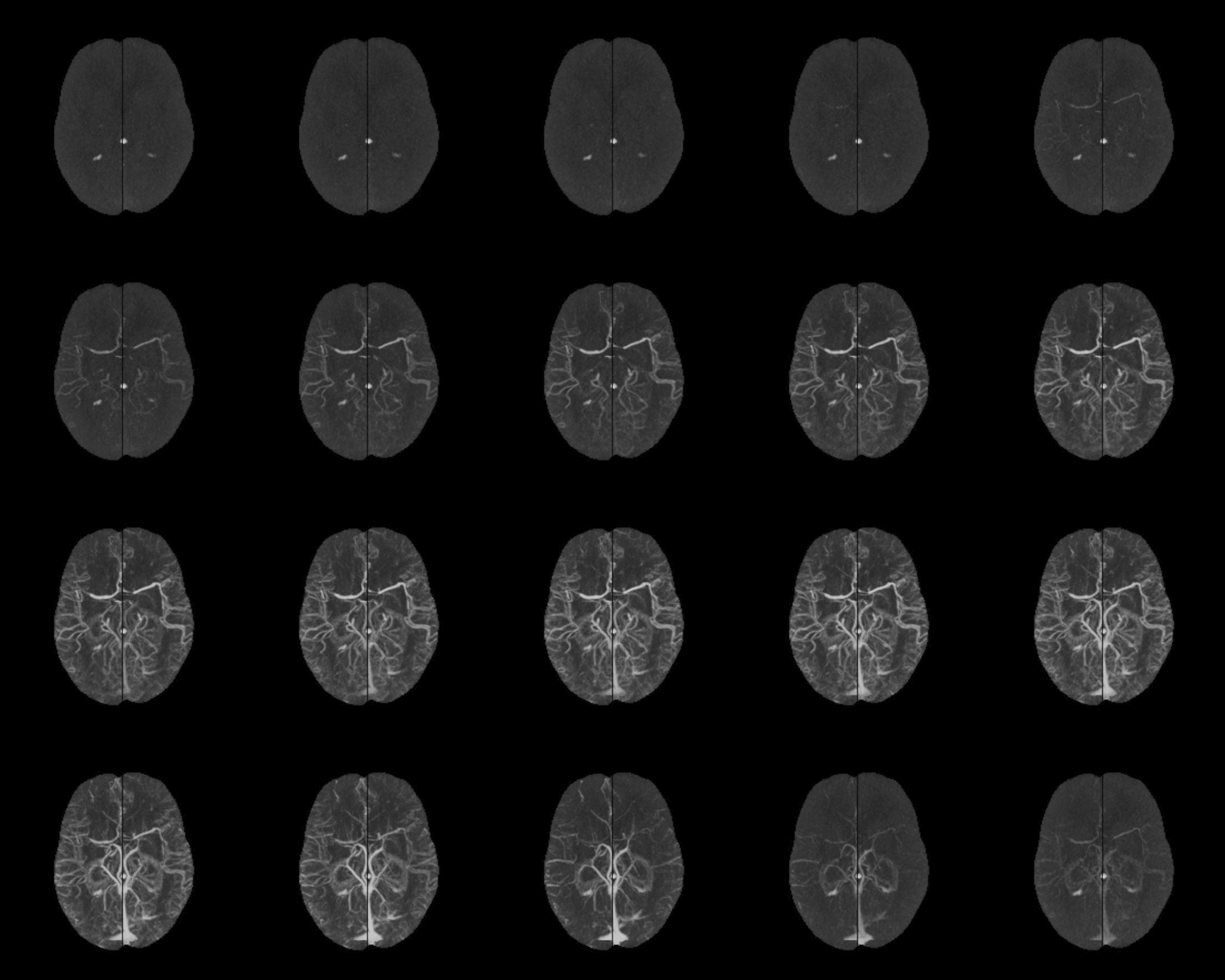
Stroke is the second leading cause of disability worldwide. In order to minimize disability, the goal of stroke treatment is to preserve tissue in the area where blood supply is decreased but sufficient to stave off cell death. Thrombectomy has been shown to offer fast and efficient reperfusion with high recanalization rates. However, due to the inherent risks of thrombectomy, indications including evidence of good collateral circulation should be present. Currently, methods for evaluating collateral circulation are limited. We are working on developing automated techniques to compute a collateral circulation score based on differences seen in mean intensities between left and right cerebral hemispheres in 4D angiography images.
Automatic Collateral Circulation Evaluation in Ischemic Stroke (ACCESS)
Sufficient collateral blood supply is crucial for favorable outcomes with endovascular treatment. The current practice of collateral scoring relies on visual inspection and thus can suffer from inter and intra-rater inconsistency. We present a robust and automatic method to score cerebral collateral blood supply to aid ischemic stroke treatment decision making.
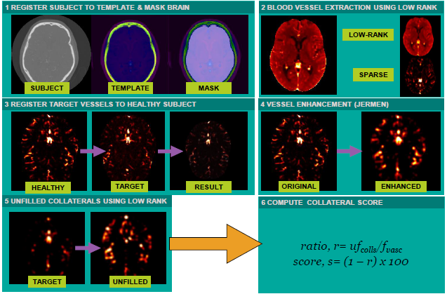
Our proposed method, ACCESS estimates a target patient’s unfilled cerebrovasculature in contrast-enhanced CTA using the lack of contrast agent due to clotting. To do so, the fast Robust Matrix Completion (fRMC) algorithm with in-face extended Frank-Wolfe optimization is applied on a cohort of healthy subjects and a target patient, to model the patient’s unfilled vessels and the estimated full vasculature as sparse and low-rank components respectively. The collateral score is computed as the ratio of the unfilled vessels to the full vasculature, mimicking existing clinical protocols. ACCESS was tested with 46 stroke patients and obtained a mean AUC of 85.39%.
ACCESS automates collateral scoring to mitigate the shortcomings of the standard clinical practice. It is a robust approach, which resembles how radiologists score clinical scans, and can be used to help radiologists in clinical decisions of stroke treatment.
Publications
- Aktar M.*, Xiao Y., Tampieri D., Rivaz, H., Kersten-Oertel, M., A Radiomics-based Machine Learning Approach to Assess Collateral Circulation in Ischemic Stroke on Non-contrast Computed Tomography. Multimodal Learning for Clinical Decision Support and Clinical Images-Based Procedures (CLIP2020). [PDF (preprint)]
- Aktar M.*, Tampieri D., Rivaz, H., Kersten-Oertel, M., Xiao Y. Automatic Collateral Circulation Scoring in Ischemic Stroke using 4D CT Angiography with Low-Rank and Sparse Matrix Decomposition. International Journal of Computer Assisted Radiology and Surgery (2020). https://doi.org/10.1007/s11548-020-02216-w
- Xiao Y, Alamer A, Fonov V, Lo BWY, Tampieri D, Collins DL, Rivaz H, Kersten-Oertel M. Towards automatic collateral circulation score evaluation in ischemic stroke using image decompositions and support vector machines. In: Cardoso M. et al. (eds) Molecular Imaging, Reconstruction and Analysis of Moving Body Organs, and Stroke Imaging and Treatment. CMMI 2017, SWITCH 2017, RAMBO 2017. Lecture Notes in Computer Science, vol 10555. Springer, Cham.
- Kersten-Oertel, M., Alamer, A., Fonov, V., Lo, B. W., Tampieri, D., & Collins, D. L. (2020). Towards a Computed Collateral Circulation Score in Ischemic Stroke. arXiv preprint arXiv:2001.07169.